How to
Search
Find
How to
Join
Play Video
Video time control bar
0:00
▶️
⏸️
🔊
Audio volume control bar
0:00
/
0:00
↘️ 0.25
↘️ 0.5
↘️ 0.75
➡️ 1
↗️ 1.25
↗️ 1.5
↗️ 1.75
↗️ 2
↔️
↕️
Timecodes:
Related videos:
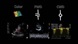
Spectral Doppler
Continuous vs Pulsed Wave Doppler Ultrasound | Ultrasound Course | Radiology Physics Course #21
Bedside Ultrasound Basic Cardiac US
Point of Care Echo: Stroke Volume Determination
E/A Ratio and Diastolic Dysfunction
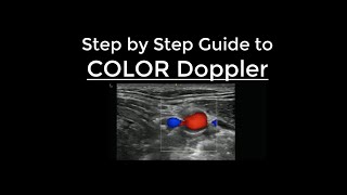
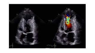
How to use Color Doppler on Ultrasound - Step by Step Guide
Pitfalls of VTI
All about TAPSE! (Echocardiography)
Diastolic Function — A Simple Approach
Ultrasound Physics Scanning Modes Pulsed Wave Doppler
Tissue Doppler Step by Step - Medial e' Example
Evaluating RVOT (Right Ventricular Outflow Tract) With Ultrasound and Doppler
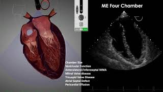
Transesophageal Echocardiography (TEE) Imaging
How to Assess the Level of Obstruction with Echocardiography in Patients with HCM
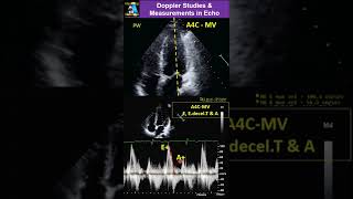
Doppler Studies & Measurements in Echo
Echocardiography Mock Exam 2 | Collection of 25 Echo Cases #cardiology #echo #echocardiography
Standardisierte Echokardiographie - Normalbefund
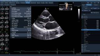
amazing LVOT VTI measurement [live]
Doppler Ultrasound Part 1 - Principles (w/ focus on Spectral Waveforms)
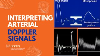
Interpreting Arterial Doppler Signals


![amazing LVOT VTI measurement [live]](https://i.ytimg.com/vi/Sl5Qmwy8dAY/mqdefault.jpg)