How to
Search
Find
How to
Join
Play Video
Video time control bar
0:00
▶️
⏸️
🔊
Audio volume control bar
0:00
/
0:00
↘️ 0.25
↘️ 0.5
↘️ 0.75
➡️ 1
↗️ 1.25
↗️ 1.5
↗️ 1.75
↗️ 2
↔️
↕️
Timecodes:
Related videos:
Cardiopulmonary POCUS 7 - Left Ventricular Failure
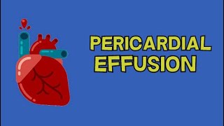
POCUS Teaching Point Pericardial effusion
Pericardial effusions and tamponade: basic POCUS evaluation
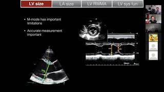
LV size & function
Staying Strong: Tips to Preserve Muscle Strength with Age.
PERICARDIAL EFFUSION
POCUS - Lung Ultrasound: Understanding B Lines and Hepatization
Pericardial Effusion and Cardiac Tamponade
Pleural Effusion Lung Ultrasound. Perioperative & Critical Care POCUS
Subcostal (subxiphoid) Echocardiography Scan Techniques
60 Seconds of Echo Teaching Question: Ventricular Septal Defect?
Differentiating pleural vs pericardial effusion by anatomy on echo
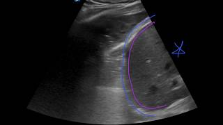
How to...Measure a Pericardial Effusion
Cardiac Arrest - ACLS Review
Left Pleural Effusion Seen on Transthoracic Echo.
Heart PLAX Moderate Pericardial Effusion
Diagnosis of SVT in the EP lab
Pleural Effusion versus Pericardial Effusion on Ultrasound Imaging
Abdominal X-Rays Made Easy
Dilated Cardiomyopathy